
Promaxo MRI-Guided Prostate Biopsy
When prostate cancer is suspected and a biopsy is recommended, the location of tissue sampling matters. At the Center for Advanced Urology, Promaxo MRI-Guided Prostate Biopsy combines MRI imaging with tissue sampling to help the physician target areas of concern identified during prostate evaluation.
A prostate biopsy is the procedure used to obtain tissue for examination by a pathologist. MRI can add information about prostate anatomy and the location of suspicious lesions, while pathology determines whether cancer is present in the sampled tissue.
Center for Advanced Urology offers Promaxo MRI-Guided Prostate Biopsy as part of an individualized diagnostic approach led by Stuart M. Diamond, MD, FACS, a board-certified urologist with more than 30 years of urologic experience.

5.0 from 550+ Reviews | Moorestown & Salem, NJ
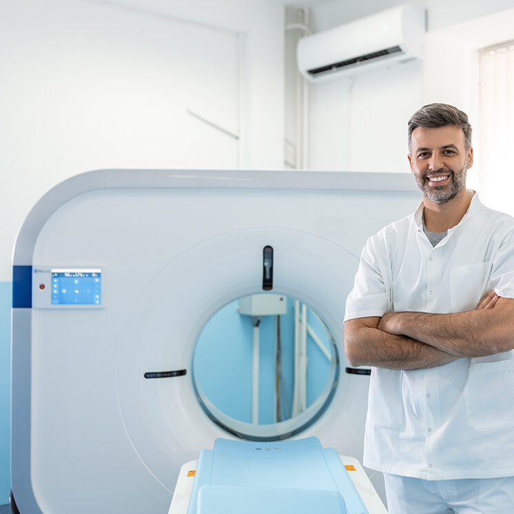
What Is a Promaxo MRI-Guided Prostate Biopsy?
A Promaxo MRI-Guided Prostate Biopsy combines MR visualization with targeted prostate tissue sampling. The Promaxo MRI System is an office-based MRI system designed to display the prostate and adjoining tissues and support MR-guided targeting of prostate lesions.
When a suspicious area has been identified, MRI information can help the physician direct the biopsy toward that region. The Promaxo system is designed so MRI guidance can be incorporated into the biopsy procedure rather than relying only on images obtained during a separate examination.
The purpose of the biopsy is to obtain tissue that can be examined under a microscope. MRI helps with targeting, but it does not replace pathology or establish a cancer diagnosis on its own.
When Is MRI-Guided Prostate Biopsy Considered?
A prostate biopsy may be considered when PSA results, physical examination findings, MRI findings, or other risk factors create enough concern to warrant tissue evaluation.
Potential reasons for biopsy include:
An elevated PSA does not by itself mean that cancer is present. PSA can rise because of benign prostate enlargement, inflammation, infection, and other conditions. Your urologist considers PSA in the context of your overall clinical picture before recommending biopsy.
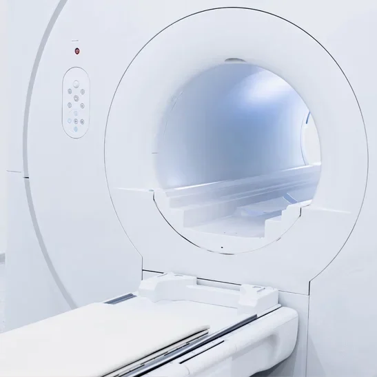
Why Perform MRI Before a Prostate Biopsy?
For men with suspected clinically localized prostate cancer, the European Association of Urology recommends performing prostate MRI before biopsy. When MRI identifies a suspicious lesion, MRI-targeted biopsy can be used to direct sampling toward that area.
MRI can therefore help answer two related questions: whether there is an area that warrants tissue evaluation and, when biopsy is appropriate, where targeted samples should be obtained.
Not every patient with a suspicious MRI requires the same biopsy strategy. The decision depends on the MRI findings, PSA density, previous biopsy history, examination findings, family history, and other clinical factors.
A systematic biopsy samples predetermined areas of the prostate according to an established sampling pattern.
A targeted biopsy uses imaging information to focus sampling on a specific area considered suspicious.
These approaches are not always mutually exclusive. Depending on the patient's circumstances and MRI findings, a physician may recommend targeted sampling together with additional systematic or perilesional samples. Current EAU guidance recognizes cognitive targeting, ultrasound/MRI fusion, and direct in-bore MRI guidance as approaches to MRI-targeted biopsy.
The appropriate strategy is determined by the patient's individual risk and the clinical information available before the procedure.
MRI guidance means that imaging is incorporated into the biopsy workflow so the physician can use MRI information while directing the procedure.
For a suspicious lesion, this can help the physician identify the relevant anatomy and guide the biopsy toward the area being evaluated. The benefit is targeted sampling based on information about where the lesion is located within the prostate.
MRI guidance does not guarantee that cancer will be found or eliminate the possibility of a false-negative biopsy. MRI can miss some cancers, and a biopsy only samples specific areas of tissue. The pathology result must therefore be interpreted together with the MRI, PSA, examination, and other clinical findings.
Before recommending an MRI-guided biopsy, your physician reviews your history and determines whether tissue sampling is appropriate.
This evaluation may include:
PSA and Related Information
Your physician may review current and previous PSA results, PSA trends, PSA density, and other laboratory information relevant to your prostate cancer risk.
Medical and Family History
The evaluation may include previous prostate conditions, prior biopsies, medications, family history of prostate cancer, genetic risk factors, and other medical conditions.
Previous Imaging and Biopsy Results
Prior prostate MRI, biopsy pathology, or other prostate testing can provide important context when determining whether another biopsy is appropriate and where additional tissue should be obtained.
MRI Findings
If prostate MRI identifies a suspicious lesion, the location and characteristics of that lesion may help determine whether targeted biopsy should be performed.
Your preparation instructions depend on the biopsy approach, your medical history, medications, and Center for Advanced Urology's procedural protocol.
Before the procedure, tell your medical team about:
- Prescription medications
- Blood thinners or antiplatelet medications
- Medication or antibiotic allergies
- Previous reactions to anesthesia or medications
- Urinary tract infections or other recent infections
- Bleeding disorders
- Significant medical conditions
- Previous prostate biopsies or procedures
Your physician may provide specific instructions regarding medications that affect bleeding risk. A urine test may also be recommended when there is concern about urinary infection. If an active infection is identified, the biopsy may need to be postponed until it has been treated.
Follow the instructions provided by your care team regarding medications, food, fluids, and activity before the procedure.
Your physician and medical team will explain the procedure before it begins, including positioning, anesthesia, imaging, and tissue sampling.
Positioning and Anesthesia
You will be positioned according to the biopsy approach being used, and the treatment area will be prepared. Local anesthesia may be used to reduce discomfort.
The exact procedural steps depend on the biopsy technique and your individual circumstances.
MRI-Guided Targeting
MRI is used to visualize the prostate and help identify the area being sampled. When a suspicious lesion is present, the physician can use the MRI information to guide the biopsy toward that target.
Tissue Sampling
Once the biopsy needle is appropriately positioned, small samples of prostate tissue are collected. The samples are then sent to a pathology laboratory for microscopic examination.
The pathology report determines whether cancer is present in the sampled tissue and provides information about the characteristics of any cancer identified.
Procedure time varies according to the biopsy approach, number of samples, imaging requirements, and individual circumstances.
Your medical team can explain what to expect on the day of your procedure, including the anticipated appointment length and any recovery instructions.
Temporary symptoms can occur after prostate biopsy. These may include:
- Mild soreness or discomfort
- Blood in the urine
- Blood in the stool, depending on the biopsy approach
- Blood in the semen
- Temporary urinary symptoms
Blood in the semen can persist for several weeks. Minor bleeding in the urine or stool may resolve sooner.
Your physician will provide individualized instructions regarding activity, medications, and follow-up.
Prostate biopsy has potential complications, including infection, bleeding, and difficulty urinating.
Contact your medical team promptly if you develop:
- Fever or chills
- Increasing or severe pain
- Significant or persistent bleeding
- Difficulty urinating
- Other symptoms that cocern you after the procedure
Follow the specific aftercare instructions provided by your physician.
Like any invasive procedure, prostate biopsy carries risks. These may include bleeding, infection, temporary urinary difficulties, soreness, and other complications requiring medical attention.
MRI itself does not use ionizing radiation. However, MRI has separate safety considerations for patients with certain implants, devices, or metallic objects. The medical team will complete appropriate MRI safety screening before the procedure.
MRI-guided targeting also has limitations. A suspicious MRI appearance does not prove cancer is present, and a biopsy may miss cancer that is outside the sampled tissue. MRI and biopsy results must therefore be interpreted as part of the broader diagnostic evaluation.
After the procedure, a pathologist examines the tissue samples under a microscope.
If No Cancer Is Found
A negative biopsy means that cancer was not identified in the tissue that was sampled. It does not automatically eliminate the need for future evaluation.
If PSA levels, MRI findings, examination results, or other factors remain concerning, your urologist may recommend continued monitoring, repeat imaging, additional testing, or another biopsy.
If Cancer Is Found
If prostate cancer is identified, the pathology report provides information about the tumor, including its grade. The grade helps describe how abnormal the cancer cells appear and contributes to determining the cancer's risk category.
Your physician will interpret the pathology findings alongside your PSA, MRI, examination, and other clinical information.
A prostate cancer diagnosis does not automatically mean that immediate treatment is necessary.
Some cancers may be appropriate for active surveillance, while others may require surgery, radiation, medication, or another treatment approach. Recommendations depend on factors such as tumor grade, disease extent, overall health, and individual circumstances.
The Center for Advanced Urology can discuss the significance of your pathology findings and the next steps in your diagnostic or treatment plan.
Promaxo MRI-Guided Prostate Biopsy in South Jersey & Philadelphia
Center for Advanced Urology provides urologic care through two South Jersey offices.
Salem
MOORESTOWN
Patients throughout South Jersey and the greater Philadelphia area can contact the practice to discuss their prostate evaluation and determine whether MRI-guided biopsy may be appropriate.

Why Choose Center for Advanced Urology?
A prostate biopsy involves more than obtaining tissue. Patient selection, clinical assessment, imaging, targeting, sampling, pathology, and interpretation all contribute to the diagnostic process.
At the Center for Advanced Urology, Stuart M. Diamond, MD, FACS, provides individualized urologic care backed by more than 30 years of experience. Dr. Diamond is a board-certified urologist and Fellow of the American College of Surgeons with experience in urology, urologic oncology, surgical care, and prostate-related conditions.
Our practice uses advanced diagnostic technology when it is clinically appropriate, including Promaxo MRI and Promaxo MRI-Guided Prostate Biopsy.
We do not treat every elevated PSA or abnormal MRI finding the same way. The appropriate diagnostic approach depends on the patient's history, risk factors, imaging, previous testing, and clinical findings.
Our Clinics
The Center for Advanced Urology provides prostate cancer evaluation and urologic care at two clinic locations, each serving the communities listed below.
Moorestown
Salem
Promaxo MRI-Guided Biopsy FAQs
It is a prostate biopsy performed with MRI guidance to help the physician target an area of concern within the prostate. Promaxo is an office-based MRI system designed to support MR-guided targeting of prostate lesions.
A biopsy may be considered when PSA, examination findings, prostate MRI, previous biopsy results, or other risk factors create sufficient concern for tissue evaluation.
Both procedures obtain tissue for pathological examination. MRI-guided biopsy adds imaging information to help target a suspicious area.
MRI can improve identification and targeting of suspicious prostate lesions, but no biopsy technique can detect every cancer. The most appropriate approach depends on the patient's MRI findings and overall clinical risk. Current EAU guidance supports MRI-targeted biopsy when a suspicious lesion is identified.
A prostate biopsy can cause temporary discomfort. Local anesthesia may be used to reduce pain during the procedure, depending on the biopsy approach.
Preparation varies. Your physician may review medications, particularly blood thinners, assess for infection, and provide instructions regarding medications, food, fluids, and activity.
Most post-biopsy symptoms are temporary. Soreness and minor bleeding can occur, while blood in the semen may continue for several weeks. Your physician will provide individualized recovery instructions.
Pathology processing takes time, and turnaround varies. Your care team will explain when you should expect the results and how they will be reviewed with you.
A negative biopsy means cancer was not identified in the tissue sampled. Your urologist may still recommend monitoring or additional evaluation if PSA, MRI, examination findings, or other risk factors remain concerning.
Your physician will review the pathology report and explain how the findings relate to your PSA, MRI, examination, and other risk factors. Depending on the cancer's characteristics, the next step may involve active surveillance, additional testing, or treatment planning.
No. MRI can miss some cancers, and biopsy evaluates only the tissue that is sampled. Your physician considers the biopsy findings together with the rest of your clinical information.
Contact Us
Discuss Your Prostate Biopsy Options
An elevated PSA, abnormal prostate examination, suspicious MRI finding, or continued concern after a previous biopsy may lead your urologist to recommend tissue evaluation.
Center for Advanced Urology offers Promaxo MRI-Guided Prostate Biopsy for appropriate patients. The procedure incorporates MRI guidance into the biopsy process to help target areas of concern identified during prostate evaluation.
Stuart M. Diamond, MD, FACS can review your individual findings and determine whether an MRI-guided prostate biopsy is appropriate for your situation.
Call Center for Advanced Urology at (856) 339-4466 to schedule a consultation.