
Promaxo MRI
When PSA results, a prostate examination, or other findings raise concern about prostate cancer, imaging can provide information that blood testing and physical examination cannot. Prostate MRI creates detailed images of the prostate and can help identify areas that may require additional evaluation.
Center for Advanced Urology offers Promaxo MRI, an advanced MRI platform designed for point-of-care imaging and MRI-guided prostate procedures. Dr. Stuart M. Diamond, MD, FACS, considers each patient's PSA history, medical history, examination findings, prostate anatomy, and other risk factors when determining whether prostate MRI is appropriate.
MRI does not diagnose prostate cancer by itself. Its findings are considered alongside the rest of your clinical evaluation to determine whether monitoring, additional testing, or prostate biopsy may be appropriate.
Center for Advanced Urology provides urologic care to patients throughout the South Jersey and Philadelphia regions

5.0 from 550+ Reviews | Moorestown & Salem, NJ
What Is Promaxo MRI?
Promaxo MRI is a compact MRI platform designed for point-of-care use and MRI-guided prostate procedures. The Promaxo MRI System has
FDA 510(k) clearance for use in MRI-guided prostate biopsy.
MRI uses magnetic fields and radiofrequency energy to create images of internal structures without ionizing radiation. For prostate evaluation, these images can show prostate anatomy and areas that appear suspicious for clinically significant prostate cancer.
The reason for imaging varies by patient. Some men undergo MRI because of an elevated or changing PSA, while others may have an abnormal prostate examination, persistent concern after a previous biopsy, or known prostate cancer.

What Can a Prostate MRI Show?
MRI can show the size and structure of the prostate and identify areas with imaging characteristics that warrant further investigation.
MRI has limitations. Inflammation and benign prostate conditions can sometimes resemble cancer, while some cancers may be difficult to detect. Image quality, the imaging protocol, and interpretation can also affect the information available from the examination.
For that reason, MRI findings are considered within the broader clinical assessment rather than in isolation.
When Is a Prostate MRI Recommended?
Prostate MRI is not necessary for every man with a prostate-related concern. Your urologist determines whether imaging can help answer the specific clinical question being investigated.
MRI may be considered for:
-
Age
The risk of prostate cancer increases with age. Prostate cancer can occur in younger men, but advancing age is an important factor when determining screening and evaluation needs.
-
Family History
A father, brother, or son with prostate cancer can increase an individual's risk. A family history of certain other cancers may also be relevant because some inherited genetic changes are associated with prostate cancer.
Tell your urologist about significant cancer diagnoses in your family.
-
Genetic Factors
Inherited changes involving genes such as BRCA1 and BRCA2, as well as changes associated with Lynch syndrome, can be linked to increased prostate cancer risk.
Men with a strong family history or other high-risk features may benefit from genetic counseling or testing.
-
Other Risk Factors
Your age, family history, previous testing, overall health, and other clinical information may all contribute to risk assessment.
Prostate MRI findings may be reported using the Prostate Imaging-Reporting and Data System (PI-RADS), a standardized system for describing the likelihood that an imaging finding represents clinically significant prostate cancer.
Scores range from 1 to 5:
- PI-RADS 1: Very low likelihood
- PI-RADS 2: Low likelihood
- PI-RADS 3: Intermediate or indeterminate
- PI-RADS 4: High likelihood
- PI-RADS 5: Very high likelihood
A PI-RADS score is not a cancer diagnosis. A higher score may lead to discussion of biopsy or additional evaluation, while a lower score may support surveillance when the rest of the clinical picture is reassuring.
Your physician considers the score together with PSA, PSA density, family history, examination findings, previous biopsy results, and other relevant factors.
No. A reassuring MRI can lower the likelihood of clinically significant prostate cancer in an appropriate clinical setting, but it cannot completely exclude cancer.
If the MRI does not show a suspicious lesion, your physician may still recommend further evaluation when other risk factors remain concerning. Depending on your circumstances, that may include PSA monitoring, additional testing, repeat imaging, or biopsy.
Preparation depends on the imaging protocol and the reason for your examination. Our team will provide instructions specific to your appointment.
Tell your medical team if you have:
- Implanted medical devices
- Metal implants or metal fragments
- Previous surgeries
- Kidney disease
- Known allergies
- Previous reactions to contrast material
- Current medications
- Significant medical conditions
- Claustrophobia or anxiety related to MRI
Because MRI uses a strong magnetic field, safety screening is required before imaging. Certain implants or metallic objects may affect whether an MRI can be performed safely.
You may also be asked to remove jewelry, watches, hearing aids, or other metallic objects before the examination.
Not every prostate MRI uses contrast material. The decision depends on the imaging protocol and the clinical purpose of the examination.
If contrast is planned, tell your medical team about kidney disease, allergies, or previous reactions to contrast material. Our imaging staff will explain whether contrast is part of your examination and whether any additional preparation is required.
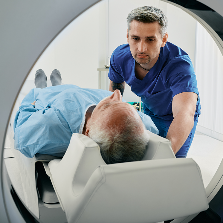
You will be positioned according to the Promaxo system and the type of examination being performed. Remaining as still as possible during image acquisition is important.
MRI equipment can produce tapping, knocking, humming, or other loud sounds, and hearing protection may be provided.
The examination itself is generally painless. Some patients may find remaining still or the imaging environment uncomfortable. If you have experienced claustrophobia or significant anxiety during previous MRI examinations, tell your medical team before the appointment.
Promaxo is designed as a compact, point-of-care MRI platform, so its physical configuration differs from the large cylindrical scanners commonly used for general imaging. Positioning and the imaging process depend on the examination being performed.
After the images have been obtained and interpreted, your physician reviews the findings alongside your other clinical information.
The next step depends on the reason for the examination and the findings.
No Suspicious Lesion
A reassuring MRI may support continued monitoring when overall risk is low. Your physician may still consider PSA levels, PSA density, family history, examination findings, and previous testing before deciding whether additional evaluation is needed.
Suspicious Lesion
A suspicious finding may lead to discussion of prostate biopsy. MRI can help localize an area of concern so tissue samples can be directed toward it when appropriate.
Center for Advanced Urology also provides Promaxo MRI-Guided Biopsy for appropriate patients. MRI-guided biopsy is a separate diagnostic procedure with its own evaluation and preparation requirements.
Previously Diagnosed Prostate Cancer
MRI findings may contribute to assessment of known prostate cancer, including evaluation of local anatomy and disease extent.
Treatment decisions involve more than imaging. Biopsy results, cancer grade and stage, overall health, and individual considerations all contribute to treatment planning.
Prostate MRI in South Jersey & Philadelphia
Center for Advanced Urology provides urologic evaluation and treatment to patients throughout the South Jersey and Philadelphia regions.
Salem
MOORESTOWN
Patients can contact our practice to determine which location is appropriate for their prostate evaluation.
Why Choose Center for Advanced Urology?

More Than 30 Years of Urologic Experience
Stuart M. Diamond, MD, FACS, is a board-certified urologist and genitourinary surgeon with more than 30 years of experience. His background includes general urology, urologic oncology, surgical care, laparoscopic and robotic surgery, clinical research, and academic medicine.

Point-of-Care Prostate Imaging
Promaxo MRI provides a compact MRI platform designed for prostate imaging and MRI-guided procedures. When clinically appropriate, the technology can contribute to decisions about further evaluation or biopsy.

Imaging Within Urologic Care
We consider prostate imaging alongside PSA history, examination findings, previous testing, medical history, and other relevant risk factors. This helps place an MRI finding in the context of the patient's overall evaluation.

Individualized Diagnostic Planning
An elevated PSA does not automatically mean that MRI or biopsy is necessary. Likewise, an abnormal MRI does not automatically establish a cancer diagnosis. We determine the appropriate next step based on the patient's clinical circumstances.
Our Clinics
The Center for Advanced Urology provides prostate evaluation and urologic care at two clinic locations, each serving the communities listed below .
Moorestown
Salem
Promaxo MRI FAQs
Promaxo MRI is a compact, point-of-care MRI platform designed for prostate imaging and MRI-guided procedures. The Promaxo MRI System has FDA 510(k) clearance for use in MRI-guided prostate biopsy.
No. MRI can identify areas that appear suspicious, but imaging alone does not establish a tissue diagnosis. A biopsy may be required to confirm prostate cancer.
MRI can identify and localize suspicious areas within the prostate. When appropriate, that information can help determine whether biopsy is warranted and where samples should be directed.
PI-RADS is a standardized prostate MRI reporting system that assigns scores from 1 to 5 according to the level of suspicion for clinically significant prostate cancer. The score does not determine whether cancer is present.
No. MRI uses magnetic fields and radiofrequency energy rather than ionizing radiation.
The imaging itself is generally painless. Some patients may find remaining still or the MRI environment uncomfortable. Tell your medical team if you have experienced claustrophobia or anxiety during previous imaging.
Not necessarily. Some prostate MRI protocols use contrast while others do not. Your imaging protocol depends on the clinical question and your physician's recommendation.
Your physician will review the finding with your other clinical information. If tissue evaluation is appropriate, a prostate biopsy may be recommended. MRI information can help direct biopsy sampling toward a suspicious lesion.
Center for Advanced Urology offers Promaxo MRI to patients across South Jersey and the greater Philadelphia area. Contact our practice to discuss your prostate evaluation and whether MRI may be appropriate.
Contact Us
Discuss Your Prostate Imaging Options
If you have an elevated PSA, an abnormal prostate examination, persistent concern after a previous biopsy, or another clinical reason to investigate your prostate, MRI may provide useful information for the next stage of evaluation.
At the Center for Advanced Urology, we consider Promaxo MRI as part of a broader physician-led assessment. Dr. Stuart M. Diamond can review your individual circumstances and determine whether prostate MRI may be appropriate and what additional evaluation may be warranted.
Call Center for Advanced Urology at (856) 339-4466 to discuss your prostate imaging options.